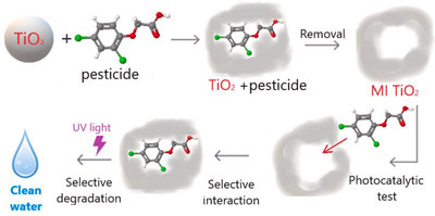
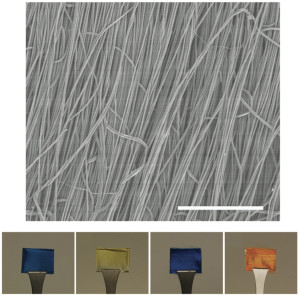
In order to achieve a selective removal of specific pesticides from water, we synthesized, through the sol–gel technique, molecularly imprinted TiO2 photocatalysts with the only use of the standard reactants of the TiO2 solgel synthesis together with the pesticide molecules, without any addition of further reactants supports or matrices. It is a new, easy, smart and scalable method that avoid the multistep and solvent–consuming procedures, typical of the molecular imprinting. Two widely–used pesticides, i.e. the herbicide 2,4D, and the insecticide imidacloprid, were chosen as template for the molecular imprinting and as contaminants target for the photocatalytic tests. A remarkable enhancement of the photocatalytic activity was verified with the TiO2 imprinted with the corresponding pesticide-target. The selectivity of the photodegradation process was verified thanks to the comparison with the degradation of pesticides not–used as template. Furthermore, the eventual toxic effects of the molecularly imprinted materials were evaluated by biological tests.
Contact person: Roberto Fiorenza, IMM c/o Università di Catania
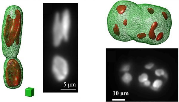
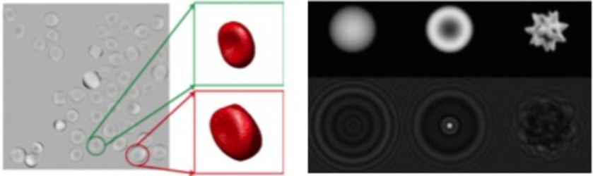
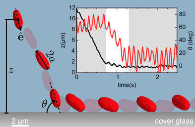 Self–propelled particles, both biological and synthetic, are stably trapped by walls and develop high concentration peaks over bounding surfaces. In swimming bacteria, like E. coli, the physical mechanism behind wall entrapment is an intricate mixture of hydrodynamic and steric interactions with a strongly anisotropic character. We have demonstrated that, by using a combination of three-axis holographic microscopy and optical tweezers, it is possible to obtain volumetric reconstructions of individual E. coli cells that are sequentially released at a controlled distance and angle from a flat solid wall. We have found that hydrodynamic couplings can slow down the cell before collision, but reorientation only occurs while the cell is in constant contact with the wall. In the trapped state, all cells swim with the average body axis pointing into the surface. The amplitude of this pitch angle is anticorrelated to the amplitude of wobbling, thus indicating that entrapment is dominated by near–field couplings between the cell body and the wall.